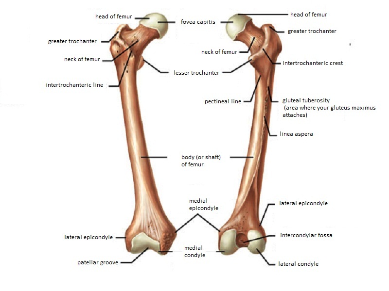
Anatomical terms of bone The medial condyle is one of the two projections on the lower extremity of femur the other being the lateral condyle. On the lateral side the primary focus of damage is within the extensor carpi radialis brevis.
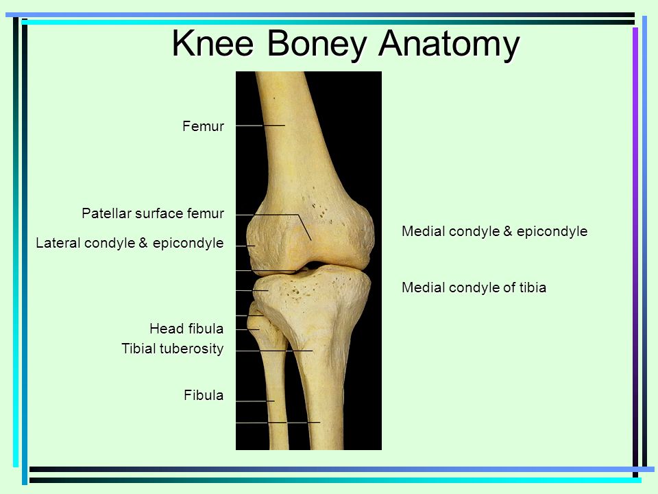
Knee Boney Anatomy Femur Medial Condyle Epicondyle Ppt Video Online Download
Isolated medial or lateral epicondyle fractures are rare.
. Artigo Original RISCOS E CONSEQUÊNCIAS DO USO DA TÉCNICA TRANSPORTAL NA RECONSTRUÇÃO DO LIGAMENTO CRUZADO ANTERIOR. Directly below it is a small depression from which a smooth well-marked groove curves obliquely upward and backward to the posterior extr. Directly below it is a small depression from which a smooth well-marked groove curves obliquely upward and backward to the posterior extremity of the condyle.
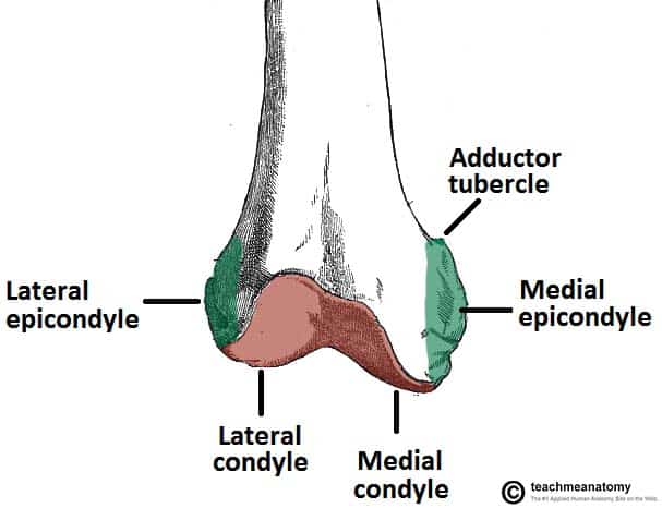
The lateral epicondyle of the femur smaller and less prominent than the medial epicondyle gives attachment to the fibular collateral ligament of the knee-joint. Talus femur tibia lateral condyle of femur medial condyle of tiba fibula medial condyle of. The femur is the only bone located within the human thigh.
Medial and lateral epicondyles ofthe femur 4. Medial epicondyle fractures. The condylar axis determined by 3D ultrasound showed good accuracy in vitro 16 SD.
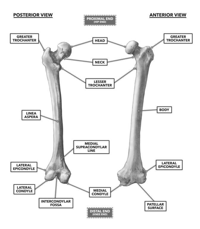
The Femur has a Greater Lessor Trochanter. They are the area of attachment of some muscles and the collateral ligaments of the knee joint. Medial and lateral epicondyles of the femur d.
Medial and lateral epicondyles Bony elevations on the non-articular areas of the condyles. Medial and Lateral Condyles. They are typically seen in children and can be challenging to identify.
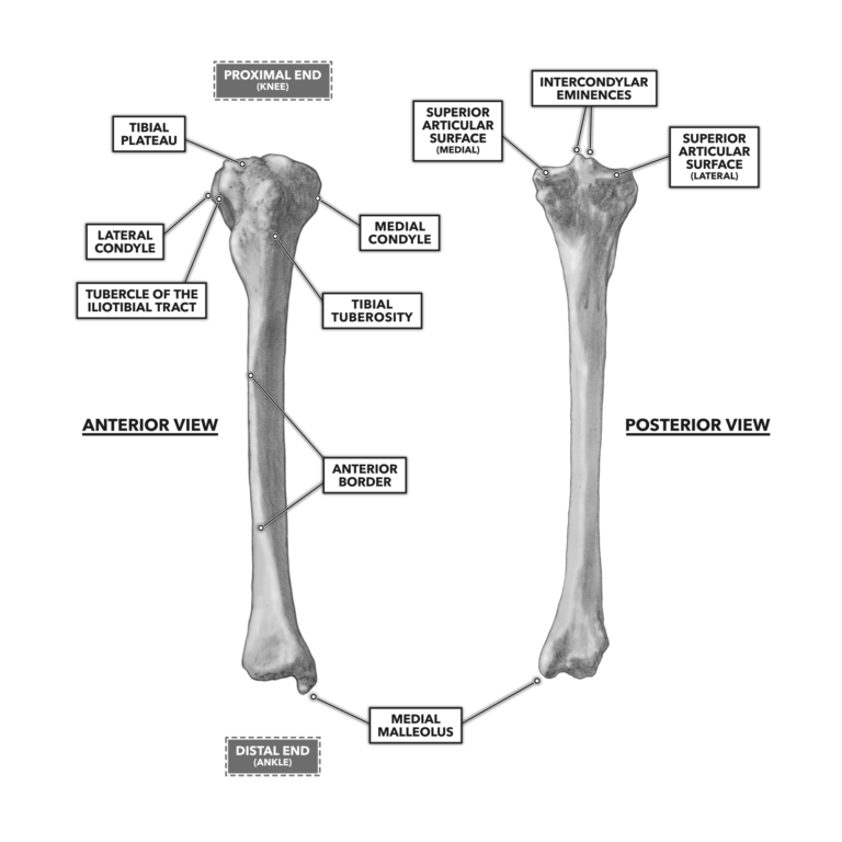
7 Does the femur have epicondyles. Styloid process of the fibula 14 Head and base of metatarsals Medial and lateral condyles of the femur. 8 Does the tibia have epicondyles.
These fractures are classified by UCPF as V3-A. Epicondylus lateralis femoris TA epicondylus lateralis ossis femoris lateral femoral tuberosity. Is the medial femoral condyle weight-bearing.
Apex of the patella b. ㅡㅡㅡㅡㅡㅡㅡㅡ Apex ofthe patella 6. 14 What is the inside of your elbow called slang.
Know the Head of the Femur and that it attaches to the hip joint called the Acetabulum. The medial condyle is larger than the lateral outer condyle due to more weight bearing caused by the centre of mass being medial to the knee. They usually form part of a more complex fracture around the femoral component.
The conventional method used markers over the femoral epicondyles. Located above the medial condyle it bears an elevation the adductor tubercle which serves for the attachment of the superficial part or tendinous insertion of the adductor magnus. Above and behind the lateral epicondyle is an area for the origin of the lateral head of the gastrocnemius above and to the medial side of which the plantaris arises.
The transepicondylar width TEW the perpendicular distance between the medial and lateral epicondyles and the distal articular surfaces DMAD DLAD and the distance between the medial and lateral epicondyles and the posterior articular surfaces PMAD DLAD were measured in 40 knees from 20 formalin-fixed adult cadavers 11 male and nine female. It also has medial and lateral epicondyles and an intercondylar fossa. The medial and lateral condyles of the tibia articulate with the a.
Intercondylar fossa A depression found on the posterior surface of the femur it lies in between the two condyles. It is both the longest and the strongest bone in the human body extending from the hip to. Greater and lesser trochanters of the femur.
The distal end of the femur features the medial and lateral condyles which articulate with the tibia and patella forming the knee joint. The medial condyle is one of the two projections on the lower extremity of femur the other being the lateral condyle. Failure to diagnose these injuries can lead to significant long term disability.
Which bone does not. The medial epicondyle of the femur is an epicondyle a bony protrusion located on the medial side of the femur at its distal end. 11 What muscles originate off the medial epicondyle.
7Tibial tuberosity 8Tibial spine 9 Ubial plateau 10Medial and lateral condyles of the tibia 11. 12 What is another name for epicondyle. 03 and good repeatability in vivo 02 RSMD.
The smaller bone that runs alongside the tibia fibula and the kneecap patella are the other bones that make the knee joint. 4 rows The medial and lateral lips unite along the middle third of the femoral shaft traveling. This tendinous part here forms an intermuscular.
9 Why is the medial epicondyle called the funny bone. RELAÇÃO ENTRE O TÚNEL FEMORAL A ARTÉRIA GENICULAR LATERAL SUPERIOR E O EPICÔNDILO LATERAL DO CÔNDILO FEMORAL RISKS AND CONSEQUENCES OF USING THE TRANSPORTAL TECHNIQUE IN. Match the structure with the appropriate name.
These are the rounded areas present at the distal end of the femur. On the medial side it is within the flexor carpi radialis and pronator teres origin. Medial epicondyle fractures represent almost all epicondyle fractures and occur when there is avulsion of the medial epicondyle.
The lateral epicondyle of the femur smaller and less prominent than the medial epicondyle gives attachment to the fibular collateral ligament of the knee-joint. The medial condyle is larger than the lateral outer condyle due to more weight bearing caused by the centre of mass being medial to the knee. Head of the fibula e.
The DynaKAD method applied to the walking calibration movement determined the medial-lateral axis closest to the ultrasound reference. 10 How many epicondyles are there. Farlex Partner Medical Dictionary Farlex 2012.
Medial and lateral condyles of the femur c. 13 What is the medial epicondyle. Lateral Medial Epicondyles Femur detailed with Head of the Femur Greater Lesser Trochanter Medial Lateral Epicondyles Single view of the Femur Bone Hip joint with Femur connection to hip joint Share.
The histology of epicondylitis has been described as angiofibroblastic hyperplasia. The articular surface of the lower end of the femur occupies the anterior inferior and posterior surfaces of the condyles. TA the epicondylus located proximal to the lateral condyle.
Lateral epicondyle of femur. A injury to any one of these structures can cause knee pain. Namely the presence of fibroblasts and vascular tissue along with degenerative and torn tendon fibers.
Crossfit Bones Of The Knee
Crossfit Bones Of The Knee
The Femur Proximal Distal Shaft Teachmeanatomy
Femur Anatomy And Attachments Bone And Spine

Leg Knee Anatomy

The Knee Musculoskeletal Key
Orif Lag Screw For Lateral Medial Femoral Epicondyle Fracture

Femur An Overview Sciencedirect Topics
0 comments
Post a Comment